Electroencephalography (EEG) utilizes standardized electrode placement, like the 10-20 system, for consistent brain activity recording. This system, detailed in guidelines by the American Electroencephalographic Society, ensures reliable data analysis.
What is Electroencephalography (EEG)?
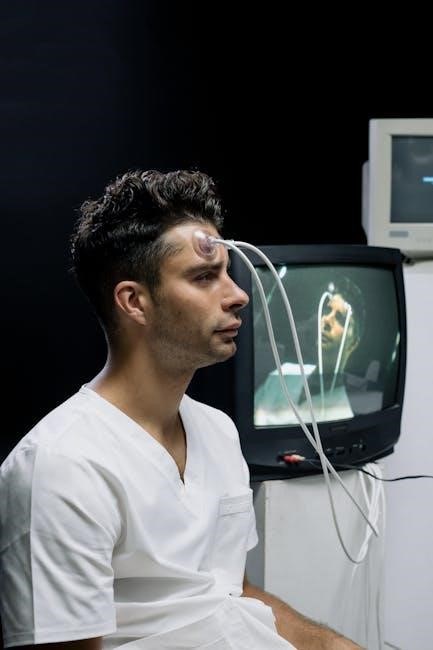
Electroencephalography (EEG) is a non-invasive neurophysiological measurement technique used to record the electrical activity of the brain. It involves placing electrodes on the scalp to detect voltage fluctuations resulting from ionic current flows within the neurons of the brain. Essentially, EEG captures the collective electrical activity of large populations of neurons.
The EEG is a crucial diagnostic tool in clinical settings, particularly for identifying and monitoring conditions like epilepsy, sleep disorders, and brain injuries. It’s also increasingly utilized in research to study cognitive processes, brain states, and neural responses. The device itself, the electroencephalogram, provides a visual representation of these brainwaves, allowing clinicians and researchers to analyze patterns and identify abnormalities.
Modern advancements, such as wireless EEG headsets like CerebAir, are expanding the possibilities for continuous and long-term brain activity monitoring, even in intensive care settings. These technologies build upon the foundational principles of EEG, relying on accurate signal detection and interpretation.
The Importance of Standardized Electrode Placement
Standardized electrode placement is paramount in Electroencephalography (EEG) to ensure data reliability and comparability across individuals and studies. Without a consistent method, variations in electrode location would introduce significant noise and hinder accurate interpretation of brain activity. The 10-20 system addresses this need by providing a universally recognized framework.
This standardization, formalized by the American Electroencephalographic Society, allows for meaningful comparisons of EEG recordings, facilitating diagnosis and research. Accurate placement, based on anatomical landmarks, minimizes artifacts and ensures that signals originate from the intended brain regions. Deviations from the standard can lead to misinterpretations and inaccurate conclusions.
Furthermore, consistent placement is vital for longitudinal studies, tracking changes in brain activity over time within the same individual. New systems like the 10-10 system and high-density EEG (hdEEG) build upon this foundation, offering increased spatial resolution while maintaining the core principle of standardized positioning.
Historical Development of the 10-20 System
The 10-20 system for EEG electrode placement didn’t emerge overnight; it was a gradual refinement born from the need for standardized brain activity mapping. Early EEG recordings lacked consistency, hindering comparative analysis. Recognizing this limitation, researchers sought a reproducible method based on readily identifiable skull measurements.
The system’s development involved defining key anatomical landmarks – the nasion, inion, and preauricular points – and establishing a percentage-based system for electrode localization. This allowed for consistent placement regardless of head size or shape. The initial guidelines were published in 1991 by the American Electroencephalographic Society, solidifying its position as the gold standard.
Over time, the 10-20 system has undergone minor revisions and expansions, leading to variations like the 10-10 system and high-density EEG (hdEEG). However, the fundamental principles of anatomical referencing and percentage-based measurements remain central, demonstrating its enduring legacy in neurophysiological research and clinical practice.

Understanding the 10-20 Electrode Placement System
EEG relies on precise electrode positioning using skull landmarks – nasion, inion, and preauricular points – and percentage-based measurements to ensure standardized, reliable recordings.
Key Anatomical Landmarks
Accurate electrode placement within the 10-20 system fundamentally depends on identifying key skull landmarks. The nasion represents the point where the nasal bone meets the frontal bone, typically located between the eyebrows. The inion is found at the lowest point of the occipital bone at the back of the head, easily palpable as a bony prominence.
Preauricular points are situated immediately in front of the ear canals, serving as crucial reference points for lateral electrode positions. These landmarks aren’t merely points; they define axes along which electrode locations are calculated. Measurements are then taken as percentages of distances between these landmarks – for instance, 10% or 20% – to determine precise electrode coordinates.
Consistent identification of these anatomical features is paramount for minimizing variability and ensuring comparability of EEG data across individuals and studies. Proper landmark localization is a foundational skill for anyone performing or interpreting EEG recordings, directly impacting the quality and reliability of the resulting brainwave analysis.
Nasion, Inion, and Preauricular Points
The nasion, a critical landmark, is the midpoint of the frontal bone’s sagital suture, easily identified between the eyebrows. Accurate nasion location establishes the anterior reference point for the 10-20 system. The inion, at the base of the skull, marks the posterior reference. Palpating the occipital bone’s lowest point reveals this key landmark, essential for posterior electrode placement.
Preauricular points, located directly anterior to the ear canals, define lateral skull dimensions. These points are crucial for establishing the transverse axis, guiding the placement of temporal and frontal electrodes. Consistent identification of these three landmarks – nasion, inion, and preauricular points – is vital for standardized EEG recordings.
Variations in skull shape necessitate careful palpation and, sometimes, visual confirmation. Precise landmark localization minimizes inter-subject variability, ensuring reliable and comparable EEG data. These anatomical references form the bedrock of the 10-20 system’s accuracy and reproducibility.
Percentage-Based Measurements
The 10-20 system employs percentage-based measurements from the nasion and inion to pinpoint electrode locations. Distances are calculated as percentages of the total head size, ensuring proportional placement regardless of individual skull dimensions. For example, electrodes are positioned at 10% and 20% of the head length from the nasion, defining frontal electrode sites.
Similarly, distances from the inion determine posterior electrode positions. Lateral measurements utilize percentages of the head width, calculated from the preauricular points. This standardized approach minimizes errors caused by variations in head size and shape, promoting consistent recordings.
Intermediate points are derived by interpolation between established landmarks. The 10-20 system’s reliance on percentages guarantees a systematic and replicable method for electrode placement. This precision is crucial for accurate brain activity mapping and reliable clinical interpretation of EEG data, facilitating comparative analyses across subjects.

Electrode Names and Locations
EEG utilizes specific electrode names – Fp1, Fp2, C3, C4, O1, O2, and others – denoting precise locations on the scalp, following the 10-20 system guidelines.
Frontal Electrodes (Fp1, Fp2, F3, F4, F7, F8)
Frontal electrodes, crucial in the 10-20 system, cover the forehead and anterior regions of the scalp. Fp1 and Fp2 are positioned prefrontally, approximately 10% and 20% of the head size from the nasion, respectively, along the midline. These electrodes are sensitive to activity in the prefrontal cortex, often linked to executive functions, attention, and personality.
F3 and F4 lie more centrally on the frontal lobe, reflecting activity from the dorsolateral prefrontal cortex, involved in working memory and cognitive control. Further laterally, F7 and F8 are positioned over the orbitofrontal cortex, associated with emotional processing and decision-making. Accurate placement, guided by anatomical landmarks like the nasion and preauricular points, is vital for reliable EEG interpretation. Variations in placement can significantly alter recorded signals, impacting clinical diagnosis and research findings. Consistent application of the 10-20 system ensures comparability across studies and patients.
Central Electrodes (C3, C4, Cz)
Central electrodes – C3, C4, and Cz – are fundamental components of the standardized 10-20 system, positioned over the central sulcus. This region is critical for processing somatosensory information and motor control. C3 represents the left hemisphere, while C4 corresponds to the right, reflecting activity related to contralateral limb movements and sensation.
Cz, the midline central electrode, serves as a key reference point and is sensitive to overall cortical activity. Placement is determined by identifying the preauricular points and measuring distances along the scalp. These electrodes are particularly valuable in detecting sensorimotor rhythms, like the mu and beta waves, which are modulated during movement and attention. Accurate placement, adhering to the 10-20 system guidelines, is essential for precise interpretation of EEG data. Deviations can lead to misidentification of brain activity and inaccurate clinical assessments. Consistent application ensures reliable and comparable results across individuals and studies.
Temporal Electrodes (T3, T4, T5, T6)
Temporal electrodes – T3, T4, T5, and T6 – within the 10-20 system, are strategically positioned over the temporal lobes. These areas are crucial for auditory processing, memory formation, and emotional regulation. T3 and T4 are located laterally, reflecting activity in the superior temporal gyri, while T5 and T6 are more posterior, covering regions involved in memory and language comprehension.

Precise placement relies on identifying key anatomical landmarks and utilizing percentage-based measurements from the nasion and preauricular points, as defined by the 10-20 system. These electrodes are particularly sensitive to temporal lobe epilepsy and can detect interictal spikes or seizure activity originating in these regions. Monitoring with temporal electrodes aids in localizing the epileptic focus. Consistent application, following standardized guidelines, is vital for accurate interpretation and reliable clinical diagnosis. Variations in placement can impact signal quality and diagnostic accuracy.
Occipital and Parietal Electrodes
Occipital (O1, O2, Oz) and Parietal (P3, P4, Pz) electrodes, within the 10-20 system, monitor visual processing and spatial awareness, crucial for comprehensive EEG analysis.
Occipital Electrodes (O1, O2, Oz)
Occipital electrodes – O1, O2, and Oz – are strategically positioned within the 10-20 system to capture electrical activity primarily originating from the occipital lobe. This region of the brain is fundamentally involved in visual processing, including the interpretation of shapes, colors, and motion. O1 is typically placed 10% of the head size behind the inion (the bony prominence at the base of the skull) on the left hemisphere, while O2 mirrors this placement on the right. Oz resides on the midline, 10% behind the inion.
These electrodes are vital for detecting alpha waves, prominent rhythmic brain activity observed during relaxed wakefulness with eyes closed. Alpha waves are often most pronounced over the occipital regions. Abnormalities in occipital electrode readings can indicate various conditions, including visual disturbances, seizures originating in the occipital lobe, or even broader neurological dysfunction. Accurate placement, adhering to the 10-20 system guidelines, is paramount for reliable interpretation of occipital lobe activity and accurate diagnosis.
Parietal Electrodes (P3, P4, Pz)
Parietal electrodes – P3, P4, and Pz – within the 10-20 system, are crucial for monitoring brain activity related to spatial orientation, navigation, and sensory integration. P3 is located 10% of the head size behind the preauricular point (a point just in front of the ear) on the left hemisphere, with P4 mirroring this position on the right. Pz sits on the midline, 10% posterior to the preauricular points.
These electrodes detect signals associated with processing tactile information, temperature, pain, and pressure. They also play a role in understanding numerical concepts and language processing. Deviations in parietal lobe activity, as observed through these electrodes, can be indicative of conditions like somatosensory deficits, spatial neglect, or seizure activity. Precise placement, following the standardized 10-20 system, is essential for accurate interpretation and reliable clinical assessment. Variations in signal patterns can help pinpoint the location and nature of neurological issues.

Reference and Ground Electrodes
Reference electrodes provide a stable baseline for EEG signals, while the ground electrode minimizes electrical interference. Proper placement, as outlined in 10-20 guidelines, is vital for data quality.
Common Reference Electrode Placements
Selecting a suitable reference electrode is crucial for accurate EEG interpretation. Several common placements are utilized within the 10-20 system, each with specific advantages and considerations. Historically, linked mastoids (A1 and A2), positioned behind the ears, served as a frequent reference point, offering a relatively stable potential. However, average reference, derived from multiple electrodes, is increasingly favored to minimize artifacts and enhance spatial resolution.
Other options include the Cz electrode, situated at the midline, and earlobes. The choice often depends on the research question and potential sources of noise. For instance, in studies focusing on frontal activity, a more posterior reference might be preferred. It’s important to note that the reference doesn’t represent brain activity itself, but rather provides a comparative baseline. Consistent reference placement across subjects is paramount for reliable group comparisons and longitudinal studies, adhering to standardized protocols like those detailed by the American Electroencephalographic Society.
The Role of the Ground Electrode
The ground electrode in EEG plays a vital safety and technical role, minimizing electrical interference and ensuring signal integrity. Typically placed on the forehead (Fpz) or mastoid process, its primary function is to provide a path of least resistance for stray electrical currents, diverting them away from the sensitive recording electrodes. This reduces noise and prevents potential hazards to the patient.
Effectively, the ground electrode creates a zero-potential reference point, stabilizing the recording system. While not directly involved in measuring brain activity, a properly functioning ground is essential for obtaining clean and reliable EEG data. Impedance checks are crucial to confirm a good connection between the ground and the patient’s skin. A high-impedance ground can introduce artifacts and compromise the quality of the recording, impacting subsequent analysis and interpretation, particularly when adhering to standardized systems like the 10-20 method.

Variations and Extensions of the 10-20 System
10-10 systems offer denser coverage, while high-density EEG (hdEEG) utilizes numerous electrodes for enhanced spatial resolution, expanding upon the foundational 10-20 method.
10-10 System
The 10-10 system represents a significant refinement of the original 10-20 system, aiming to provide a more detailed and precise mapping of the scalp’s electrical activity. Unlike the 10-20 system, which relies on measurements based on percentages of head size, the 10-10 system utilizes a 10% interpolation between electrodes. This denser electrode array allows for improved spatial resolution, capturing more nuanced brain signals.
Essentially, the 10-10 system divides the scalp into smaller, more defined areas, enabling researchers and clinicians to pinpoint the sources of brain activity with greater accuracy. This is particularly valuable in studies focusing on localized brain functions or in identifying subtle abnormalities in brainwave patterns. While the 10-20 system remains a standard for routine EEG recordings, the 10-10 system is frequently employed in research settings and specialized clinical applications where high spatial resolution is paramount. It builds upon the established landmarks of the 10-20 system – Nasion, Inion, and Preauricular points – but adds more intermediate electrodes for a more comprehensive assessment.
High-Density EEG (hdEEG)
High-Density EEG (hdEEG) represents a substantial leap beyond both the 10-20 and 10-10 systems, employing a significantly larger number of electrodes – often exceeding 256 – distributed across the scalp. This dramatic increase in electrode density allows for exceptionally detailed spatial resolution of brain activity, enabling researchers to investigate complex neural processes with unprecedented precision. Unlike traditional systems relying on fixed percentages, hdEEG often utilizes sophisticated digitization and modeling techniques to optimize electrode placement.
HdEEG is particularly valuable for source localization, attempting to pinpoint the precise origins of electrical signals within the brain. It’s frequently used in cognitive neuroscience research, investigating phenomena like language processing, memory, and attention. While requiring more complex setup and analysis, hdEEG provides a richer dataset, facilitating more accurate and nuanced interpretations of brain function. The data generated demands advanced computational methods for processing and visualization, moving beyond the simpler analyses typically associated with standard EEG recordings. It’s a powerful tool for exploring the intricacies of the human brain.

Practical Considerations for Electrode Application
Skin preparation and minimizing electrode impedance are crucial for quality EEG signals. Accurate electrode placement, following the 10-20 system, ensures reliable data collection.
Skin Preparation
Optimal skin preparation is paramount for acquiring high-quality EEG recordings. The scalp’s surface must be cleaned to remove oils, dead skin cells, and any lingering hair products that could impede electrical conductivity. Begin by gently abrading the skin with a mild abrasive, like a specialized EEG scrub or a light exfoliant, to enhance electrode-to-scalp contact.
Following abrasion, thoroughly clean the targeted areas with an alcohol-based solution – isopropyl alcohol is commonly used – to eliminate residual debris and disinfect the skin. Ensure the alcohol is completely evaporated before proceeding with electrode application, as residual alcohol can also increase impedance.
For individuals with excessive scalp oils or hair, a degreasing agent might be necessary prior to abrasion. Careful attention should be paid to areas prone to sweating, as moisture can significantly affect signal quality. Proper skin preparation dramatically reduces impedance and minimizes artifacts, ultimately leading to more reliable and interpretable EEG data.
Electrode Impedance
Electrode impedance represents the opposition to the flow of electrical current between the electrode and the scalp. Maintaining low impedance – typically below 5 kΩ, and ideally below 1 kΩ – is crucial for acquiring a clean and accurate EEG signal. High impedance introduces noise and attenuates the true brainwave activity, distorting the recording.
Impedance is influenced by several factors, including skin preparation, electrode quality, and the contact between the electrode and the scalp. Regularly check impedance levels throughout the recording session using the EEG system’s built-in impedance meter.

If impedance is too high, re-prepare the skin at that location, ensuring thorough cleaning and abrasion. Adjust electrode placement slightly or apply more conductive gel. Consistent monitoring and adjustment of impedance are essential for minimizing artifacts and maximizing the signal-to-noise ratio, ultimately ensuring the reliability of the EEG data obtained.
Ensuring Accurate Placement
Accurate electrode placement, adhering strictly to the 10-20 system guidelines, is paramount for meaningful EEG interpretation. Deviations can lead to misidentification of brain activity sources and inaccurate diagnoses. Utilize a standardized head measurement technique, starting with nasion, inion, and preauricular points, to establish precise locations.
Double-check electrode positions against anatomical landmarks and reference diagrams before commencing recording. Consider using a head measurement device or specialized software to aid in precise placement, especially for high-density EEG (hdEEG) setups.
Consistent application across patients is vital for comparative studies. Document any minor adjustments made to standard positions. Thorough training of personnel performing electrode application is essential to minimize errors and maintain data quality. Regular quality control checks and adherence to established protocols are key to reliable EEG recordings.